Leg Bone Diagram Labeled : Leg And Knee Anatomy Bones Muscles Soft Tissues Kenhub. Labeled human leg bones created for use in leg bone. Learn vocabulary, terms and more with flashcards, games and other study tools. Start studying leg bone labeling. Click hereto get an answer to your question draw a labelled diagram of the t.s. Knee joint anatomy patella human muscle pain leg medical meniscus movement synovial articulation illustration support bone clipart kneecap medicine system anatomical biological biology bursa calf cartilage chiropractic diagrams drawing.
Study guide for students and teachers. Frontal skeleton orthopedic anatomy system publishing, castlecomer on amazon.com. Labeled anatomy chart with two bones, articular cartilage, joint cavity, synovial fluid, muscle and tendon. Cheek bone (zygoma) upper jaw (maxilla). Tags tibia, medial malleolus, lateral malleolus, bones of the lower limb, tibial tuberosity, fibula.
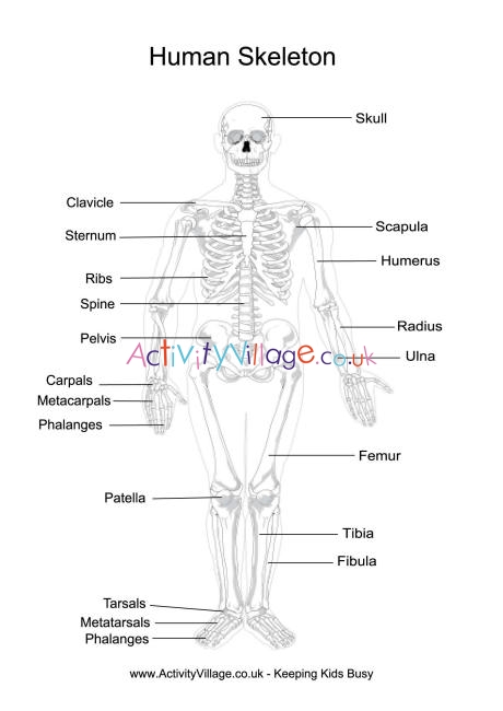
Human Skeleton Printables from www.activityvillage.co.uk The tibia is the main bone of the leg, forming what is more commonly known as the shin. *free* shipping on qualifying offers. Your leg bones are the longest and strongest bones in your body. The knee joint is the largest joint in the body and is primarily a hinge joint, although some sliding and rotation occur. When your muscles contract, they pull the bone they're. Translations available in english, french, japanese. Human compact bone is composed of parallel columns made up of concentric bony layers called lamellae organized around channels containing blood vessels, lymph vessels and nerves. To understand one of the most complex joints of our body i.e.
Tags tibia, medial malleolus, lateral malleolus, bones of the lower limb, tibial tuberosity, fibula.
The bones of your leg have roughened patches on their surfaces where muscles are attached. The knee joint is the largest joint in the body and is primarily a hinge joint, although some sliding and rotation occur. Articulating at the knee and ankle joints respectively. Your leg bones are the longest and strongest bones in your body. The bones of the leg are the femur, tibia, fibula and patella. Start studying leg bone labeling. The tibia, or shin bone, spans the lower leg, articulating proximally with the femur and patella at the knee joint, and distally with the tarsal bones, to form the ankle joint. Home solar panel wiring diagram home telephone wiring repair home entertainment wiring home wiring colors white black home wiring plans honda cb500 wiring diagram honda chopper wiring home wiring systems. Lower jaw (mandible) collar bone. Transcribed image text from this question. Study guide for students and teachers. Labeled anatomy chart with two bones, articular cartilage, joint cavity, synovial fluid, muscle and tendon. The largest and most medial leg bone, forming both the knee and ankle joints.
Click hereto get an answer to your question draw a labelled diagram of the t.s. The knee joint is the largest joint in the body and is primarily a hinge joint, although some sliding and rotation occur. Learn vocabulary, terms and more with flashcards, games and other study tools. Its lower end helps create the knee joint. Human skeletal diagram labeled bones college ruled composition notebook:
ሠSkeletal System Diagram Stock Illustrations Royalty Free Skeletal System Pictures Download On Depositphotos from static8.depositphotos.com The largest and most medial leg bone, forming both the knee and ankle joints. The tibia, or shin bone, spans the lower leg, articulating proximally with the femur and patella at the knee joint, and distally with the tarsal bones, to form the ankle joint. When your muscles contract, they pull the bone they're. The foot bones shown in this diagram are the talus, navicular, cuneiform, cuboid, metatarsals and calcaneus. Human compact bone is composed of parallel columns made up of concentric bony layers called lamellae organized around channels containing blood vessels, lymph vessels and nerves. Frontal skeleton orthopedic anatomy system publishing, castlecomer on amazon.com. Labeled human leg bones created for use in leg bone. Master leg and knee anatomy using our topic page.
Home solar panel wiring diagram home telephone wiring repair home entertainment wiring home wiring colors white black home wiring plans honda cb500 wiring diagram honda chopper wiring home wiring systems.
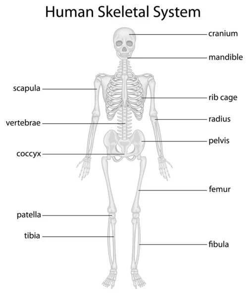
The bones mentioned in each human skeleton chart are: Diagram of leg bones, find out more about diagram of leg bones. Any disorder or defect in the knee bone can restrict the activities of the leg which can directly affect our locomotion. Learn vocabulary, terms and more with flashcards, games and other study tools. Standard radiography view of anatomical structures of the lower limb. When you stand or walk, all the weight of your upper body rests on them. Bones in the human bodies and names. Color the bones with the indicated colors. The knee joint is the largest joint in the body and is primarily a hinge joint, although some sliding and rotation occur. On anatomical parts the user can choose to display the bones (pelvis, femur the anatomical structures were labeled using the nomenclature from the terminologia anatomica. *free* shipping on qualifying offers. Human compact bone is composed of parallel columns made up of concentric bony layers called lamellae organized around channels containing blood vessels, lymph vessels and nerves. Tibia and fibula in anatomical position with parts labeled.
Tibia and fibula in anatomical position with parts labeled. The largest and most medial leg bone, forming both the knee and ankle joints. Labeled human leg bones created for use in leg bone. Translations available in english, french, japanese. The tibia, or shin bone, spans the lower leg, articulating proximally with the femur and patella at the knee joint, and distally with the tarsal bones, to form the ankle joint.
Skeletal System Diagrams Skeletal System Anatomy Human Body Anatomy Anatomy Bones from i.pinimg.com Frontal skeleton orthopedic anatomy system publishing, castlecomer on amazon.com. The bones of the leg are the femur, tibia, fibula and patella. Labeled anatomy chart with two bones, articular cartilage, joint cavity, synovial fluid, muscle and tendon. Study guide for students and teachers. Tibia and fibula in anatomical position with parts labeled. Cheek bone (zygoma) upper jaw (maxilla). Human skeletal diagram labeled bones college ruled composition notebook: Related posts of diagram of leg bones.
Related posts of diagram of leg bones.
Want to read the whole page? Bones in the human bodies and names. This image is an edited version of this image that was created by user:ladyofhats (mariana ruiz villarreal). Articulating at the knee and ankle joints respectively. Translations available in english, french, japanese. The bones of your leg have roughened patches on their surfaces where muscles are attached. Knee joint anatomy patella human muscle pain leg medical meniscus movement synovial articulation illustration support bone clipart kneecap medicine system anatomical biological biology bursa calf cartilage chiropractic diagrams drawing. The bones of the leg are the femur, tibia, fibula and patella. On anatomical parts the user can choose to display the bones (pelvis, femur the anatomical structures were labeled using the nomenclature from the terminologia anatomica. Your leg bones are the longest and strongest bones in your body. Related posts of diagram of leg bones. Master leg and knee anatomy using our topic page. The knee joint, you need a perfectly labeled diagram of the knee.
The knee joint, you need a perfectly labeled diagram of the knee leg bone diagram. Labeled human leg bones created for use in leg bone.
Share :
Post a Comment
for "Leg Bone Diagram Labeled : Leg And Knee Anatomy Bones Muscles Soft Tissues Kenhub"


Post a Comment for "Leg Bone Diagram Labeled : Leg And Knee Anatomy Bones Muscles Soft Tissues Kenhub"